Description
  1. Scout imaging as per LV function module
  2. Appropriate vendor recommended optimum T1-mapping sequence (e.g. MOLLI/ShMOLLI/SASHA etc.)
  3. SA images - single mid-ventricular slice or 3 slices planned using “3 of 5” technique
  4. Gd injection (0.1 – 0.2mmol/kg)
  5. Wait at least 15 min post contrast injection to acquire post-contrast T1.
  6. Take full blood count for haematocrit to calculate extracellular volume (ECV)
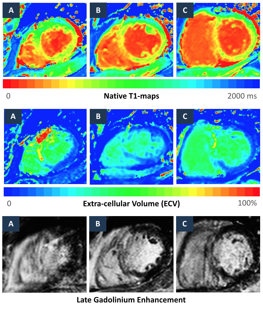
Apical (A), mid-ventricular (B) and basal (C) Native T1-maps, ECV-maps and corresponding LGE imaging for a patient with large septal infarct.

Tips and Tricks
  1. Acquire one slice per breath hold
  2. Native T1 values are reproducible but they vary by magnet strength (with 3T resulting in longer native T1 times), vendor platforms and the mapping sequence employed during acquisition
  3. Check scanner generated native T1 map for breathing artefact. Consider changing voxel size, fold-over or adding SENSE to reduce breath-hold.
  4. FOV and voxel size should not be changed between pre/post contrast T1 acquisition to allow various software to generate ECV maps