General
Echocardiography remains the first line imaging modality for the assessment of valvular heart disease.

Protocol
  1. Anatomy / LV function / RV function (RV function) module (A)
  2. Optimized cine views (B) for MV:
    • Slice thickness 5mm - consider overlapping
    • Standard long-axis views - 4CH, 2CH, 3CH
    • 3CH cine stack (perpendicular to the commissure)


  3. Phase contrast velocity encoded module (F) – AV, (MV - unreliable)
Anatomy
  1. MV components
    • Leaflets (anterior and posterior)
    • Anterolateral and posteromedial commissure
    • Annulus (oval and saddle shape)
    • Sub-valvular apparatus (chordae and papillary muscles)
  2. Mitral valve leaflets
    • Anterior leaflet: 3 scallops A1, A2, and A3 (tongue shaped)
    • Posterior leaflet: 3 scallops P1, P2, and P3 (crescent-shaped)
    • A1 / P1 located near to the LAA (antero-lateral)
    • A3 / P3 located near to the RV (postero-medial)
Report
  1. Dimensions, mass (corrected for BSA) and function
    • LV: EDV, ESV, SV, EF, mass
    • (RV EDV, ESV, SV, EF)
  2. Mitral valve morphology
    • Leaflets: thickened, restricted, tethered, bowing, prolapse, or flail
    • Annulus: thickened, fibrosis on LGE
    • Sub-valvular apparatus : thickened, restrictive, rupture
  3. Mitral regurgitation
    • MV regurgitation volume (RegV)
    • MV regurgitation fraction (RegF)
  4. Mitral stenosis
    • MV orifice area planimetry (cm²)
    • MV pressure gradients unreliable because of motion of MV apparatus
Mitral regurgitation

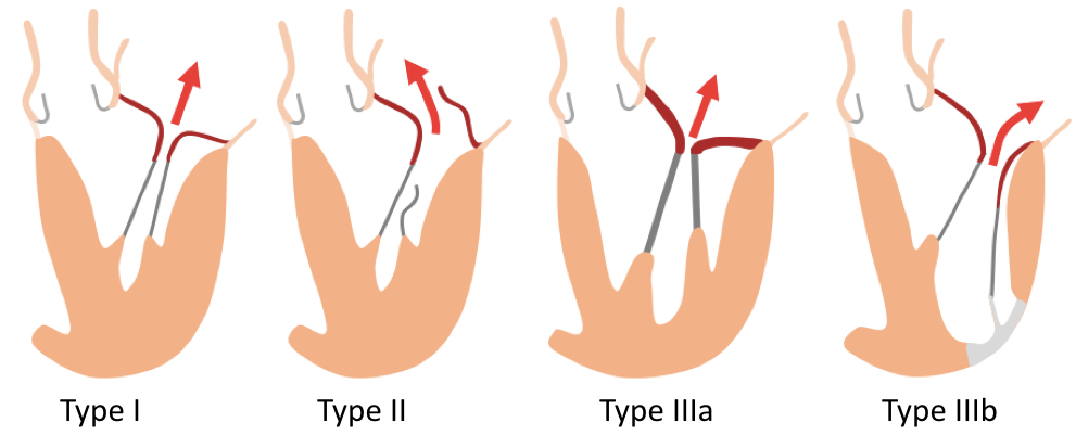
Functional classification by Carpentier

  • Type I: Normal leaflet motion (annular dilatation, leaflet perforation)
  • Type II: Excess leaflet motion (prolapse, flail, papillary muscle rupture)
  • Type IIIa: Restricted leaflet motion during systole and diastole (thickening, retraction)
  • Type IIIb: Restricted leaflet motion during systole only (tethering)

Degenerative (primary) mitral regurgitation
Caused by a primary abnormality of components of the valve apparatus
  • Chordae tendineae rupture (fibroelastic deficiency: FED)
  • Myxomatous degeneration with prolapse and/or flail (FED +, forme fruste, Barlow's disease)
  • Rheumatic heart disease
  • Endocarditis
  • Papillary muscle rupture (eg. post MI)
  • Mitral annular calcification

Functional (secondary) mitral regurgitation
Occurs despite a structurally normal mitral valve
  • Ischemic cardiomyopathy
  • Dilated cardiomyopathy
  • Hypertrophic cardiomyopathy
  • Severe left atrial dilation
Quantification
  • RegV (ml) = LV SV – SV from phase contrast VENC above AV
    (not valid in the presence of a VSD)
  • RegV (ml) alternatively = LV SV– RV SV
    (only valid in single valve disease)
  • RegF (%) = RegV / LV SV x 100
Tips and Tricks
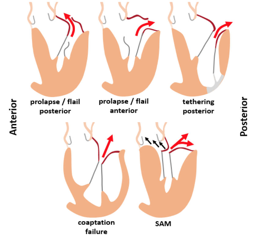
  1. Jet direction
    • Central (coaptation failure, symmetric tethering)
    • Eccentric (prolapse, flail, asymmetric tethering, SAM)
      • Anterior: prolapse / flail posterior, tethering anterior
      • Posterior: prolapse / flail anterior, tethering posterior


  2. Dilated LV suggests decompensation secondary to MR or vice versa
  3. Dilated LA suggests chronic mitral valve regurgitation
  4. Round MV annulus (diameter on 3CH and commissural 2CH view similar in size) suggests severely dilated annulus due either to LV or to LA dilatation
  5. Tethered MV leaflets
    • Asymmetric: most common posteriorly due to inferolateral MI affecting the papillary muscle
    • Symmetric: severely dilated LV or secondary to rheumatic fever
  6. RegV of >55 mL and RegF >40% predict symptom progression and other indications for surgery
  7. There is a strong correlation between post-surgical LV remodeling and MR severity as assessed by MRI
  8. Consider "automatic valve tracking“ (if available)
Mitral stenosis

Causes
  • Rheumatic valve disease
  • Systemic lupus erythematous
  • Degenerative
  • Congenital
Quantification
  • MV orifice area planimetry (cm²)
  • (MV pressure gradients unreliable because of motion of MV apparatus)
Tips and Tricks
  1. Rheumatic MS: fusion and thickening at the tips of the leaflets results in ‘doming’ of the MV leaflets (‘hockey-stick’).
  2. Degenerative MS: Annular thickening and calcification affect the base and body of MV leaflet while sparing the tips
  3. Progressive LA dilatation is marker of increased LV filling pressure and worsening MS
  4. Advanced MS has adverse impact on RV systolic function (secondary to pulmonary hypertension)
Limitiations
  • Degree of regurgitation or stenosis – cine imaging
    • Visual assessment from cine images alone is NOT recommended due to a signal void in turbulent flow
  • Flow velocity encoding – regurgitation volume / fraction
    • VENC tends to underestimate velocities due to partial volume averaging / slice orientation NOT perpendicular to the flow
    • Consider volume shift through moving aorta during cardiac circle
    • Consider regular back-flow into the coronary arteries
  • Valve area – planimetry
    • Correct imaging planes at the tip of the leaflets are fundamental
    • Note that a perfect 2D image plane of a 3D structure is impossible
References
  • Gelfand E V, Hughes S, Hauser TH, et al. Severity of mitral and aortic regurgitation as assessed by cardiovascular magnetic resonance: optimizing correlation with Doppler echocardiography. J Cardiovasc Magn Reson. 2006 Jan;8(3):503–7.
  • Djavidani B, Debl K, Lenhart M, Seitz J, et al.. Planimetry of mitral valve stenosis by magnetic resonance imaging. J Am Coll Cardiol. 2005 Jun 21;45(12):2048–53.
  • Aplin M, Kyhl K, Bjerre J, et al. Cardiac Remodelling and Function in Primary Mitral Valve Insufficiency Studied by Magnetic Resonance Imaging. Eur Heart J: Cardiovasc Imaging. 2016; (in press).
  • Adams DH, Rosenhek R, Falk V. Degenerative mitral valve regurgitation: best practice revolution. Eur Heart J. 2010 Aug;31(16):1958-66.
  • Myerson SG, d'Arcy J, Christiansen JP, et al. Determination of Clinical Outcome in Mitral Regurgitation With Cardiovascular Magnetic Resonance Quantification. Circulation. 2016 Jun 7;133(23):2287-96.
  • Uretsky S, Gillam L, Lang R, et al. Discordance between echocardiography and MRI in the assessment of mitral regurgitation severity: a prospective multicenter trial. J Am Coll Cardiol. 2015 Mar 24;65(11):1078-88.