General
  • LGE is not disease specific and can be caused by ischaemic and non-ischaemic necrosis/fibrosis, inflammatory or infectious pathology or tumourous lesions.
  • Gadolinium agents do not penetrate intact myocytes, they accumulate within the increased extracellular space or into ruptured myocytes.
  • Left: normal myocardium, center: necrosis / fibrosis with increased extracellular space, right: Acute myocardial infarction.
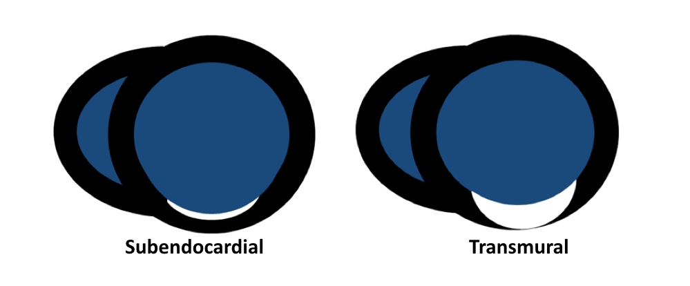
Coronary pattern Non-coronary pattern
  • From subendocardium to subepicardium (ischaemic wave front) with increasing coronary occlusion time
  • Subendocardium is typically spared
  • Circumferential extent related to size of perfusion bed and occluded coronary artery
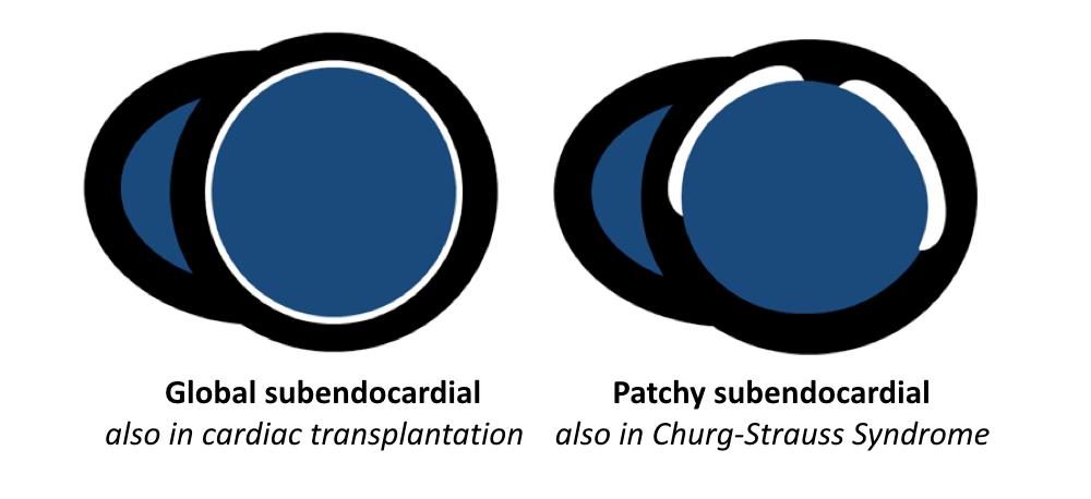
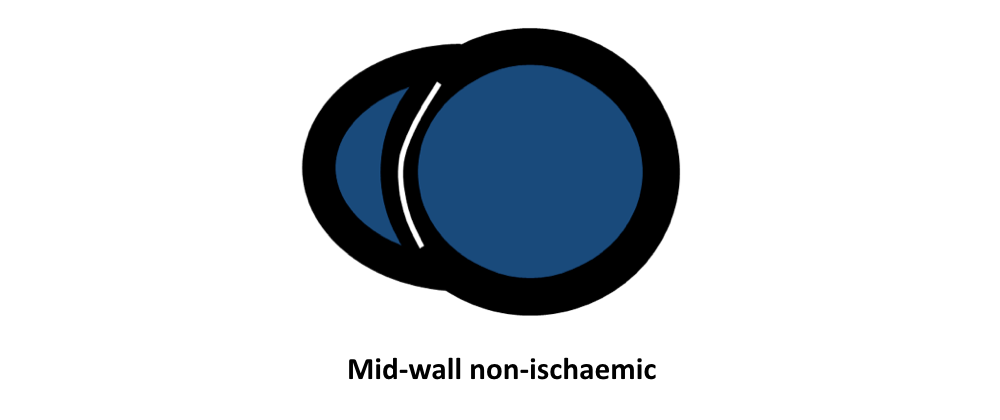
  • Often more patchy distribution, localized in mid-wall and subepicardium
  • Acute reperfused MI can be associated with MVO
  • Global subendocardial LGE (uncommon even with diffuse CAD)

Coronary pattern
  1. Subendocardial infarct
    • LGE transmurality correlates with likelihood of recovery after revascularization
    • LGE identifies arrhythmogenic substrate (mapping for ablation)
  2. Transmural myocardial infarct
    • Degree of transmurality correlates inversely with viability
  3. Ischaemic cardiomyopathy
    • Often also LV dilatation, differential diagnosis of DCM
    • Coronary LGE pattern with subendocardial/transmural LGE
    • LGE identifies arrhythmogenic substrate (mapping for ablation)
Non-coronary pattern
  1. Cardiac amyloidosis
    • Difficulty in nulling
    • Global subendocardial distribution (non-coronary pattern) or patchy subendocardial or transmural LGE
    • Base-apex LGE gradient, blood pool early darkening
    • DD AL vs. ATTR amyloidosis
      • AL: less extensive LGE, often (global) subendocardial pattern, QALE score < 13
      • ATTR: more extensive and diffuse LGE, often more diffuse and transmural pattern, QALE score ≥ 13
  2. Dilated cardiomyopathy
    • Pattern of fibrosis is related to underlying aetiology of DCM
    • In most cases lack of LGE -> consider T1 mapping
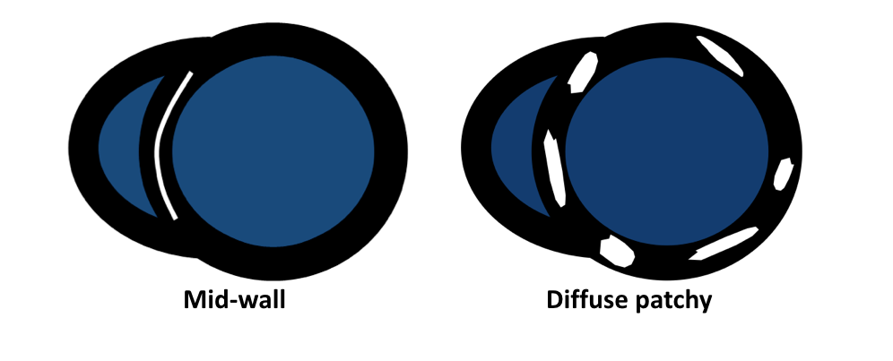
    • Diffuse mid-wall or subepicardial septal LGE (reflecting fibrosis and not matching a coronary perfusion bed)
    • Often localized LGE of the inferoseptal wall
  3. Hypertrophic cardiomyopathy
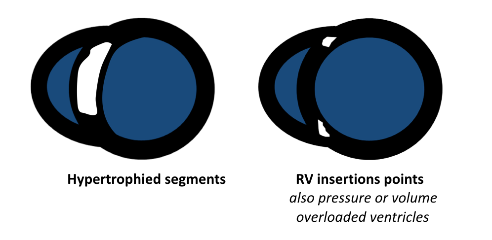
    • Focal (mid-wall) LGE of anterior and posterior RV insertion points (hinge points) and of hypertrophied segments
    • LGE in HCM indicates replacement fibrosis but may also indicate necrosis/scarring
    • Apical HCM: apical wall may develop ischaemia with wall thinning → LGE can be transmural (mimicking ischaemic injury in the absence of epicardial CAD)
  4. Systemic Sclerosis
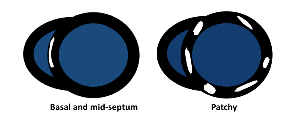
    • Basal to mid-interventricular septum and RV insertion points or diffuse patchy LGE
  5. Myocarditis
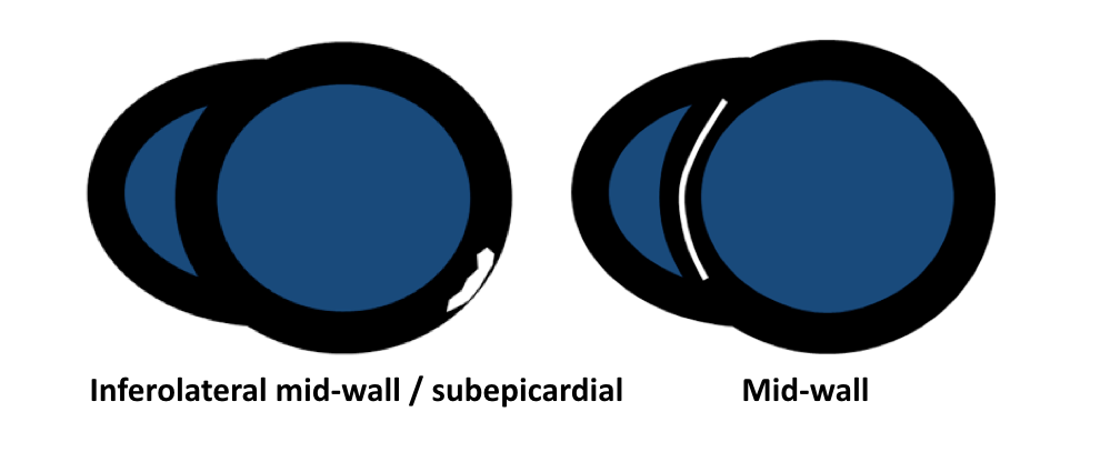
    • Lateral, typically inferolateral or inferior wall with a mid-wall to subepicardial enhancement
    • Atypical cases of myocarditis with transmural or diffuse LGE
    • Often concomitant LGE involvement of the pericardium
  6. Anderson-Fabry Disease
    • Mid-wall (or subepicardial) LGE of the inferolateral wall of mid to basal LV
    • Consider T1-Mapping: low ECV and low native T1 value of septum
  7. Sarcoidosis
    • Basal and mid-interventricular septum but also patchy LGE
  8. ARVC
    • Diffuse LGE of RV (and occasionally LV) wall
  9. Endomyocardial fibrosis (Loeffler’s endocarditis)
    • Restrictive CMP with endocardial fibrotic tissue deposition of inflow tract and apex of one or both ventricles
    • “V sign” of LGE pattern at ventricular apex, characterized by a 3-layer appearance of dark myocardium, bright thickened fibrotic endocardium, and overlying dark thrombus
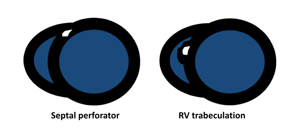
False positive LGE
  • Septal perforator (mimicking septal basal mid-wall enhancement)
  • RV trabeculation (contrast extending underneath the muscle mimicking septal enhancement)
References
  • Friedrich MG, Sechtem U, Schulz-Menger J, et al. International Consensus Group on Cardiovascular Magnetic Resonance in Myocarditis. Cardiovascular magnetic resonance in myocarditis: A JACC White Paper. J Am Coll Cardiol. 2009 Apr 28; 53(17):1475-87.
  • Vermes E, Carbone I, Friedrich MG, Merchant N. Patterns of myocardial late enhancement: typical and atypical features. Arch Cardiovasc Dis. 2012 May; 105(5):300-8.
  • Satoh H, Sano M, Suwa K, et al. Distribution of late gadolinium enhancement in various types of cardiomyopathies: Significance in differential diagnosis, clinical features and prognosis. World J Cardiol. 2014 Jul 26; 6(7):585-601.