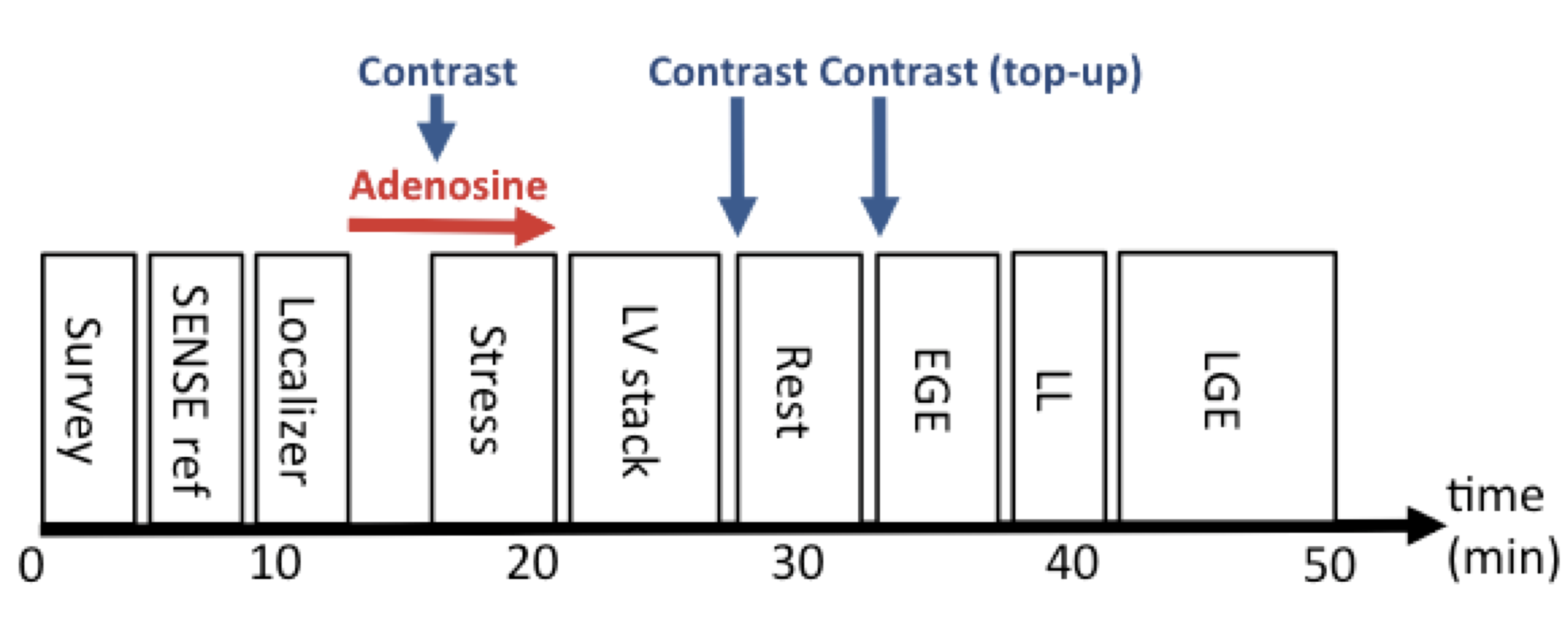
Protocol
  1. Anatomy module (A)
  2. Myocardial perfusion module (D) “dummy“
  3. Myocardial perfusion module (D) STRESS
  4. LV function module (B)
  5. Myocardial perfusion module (D) REST
  6. LGE module (E)
Report
  1. Dimensions (corrected for BSA) and function
    • LV: EDV, ESV, SV, EF / RV: EDV, ESV, SV, EF
    • Regional wall motion abnormalities (17 segments)
  2. Presence and transmural extent of scar
    • ≤25%, 26-50%, 51-75%, 76-100%
  3. Presence and transmural extent of inducible perfusion defect
  4. Correlation between scar and perfusion defect
  5. Comment on suitability of revascularization based on ischemia and viable myocardium
  6. (Presence and location of artefacts)
Key Issues
  1. Check BP /monitor ECG during adenosine perfusion
  2. Adenosine dose:
    • 140mcg/kg/min
    • Consider 170 or 210mcg/kg/min, if hemodynamic response is inadequate or after caffeine intake
  3. Contraindications for adenosine: known hypersensitivity, 2nd /3rd AV nodal block, severe reversible airways disease
Tips and Tricks
  1. Use “3 out of 5” technique to position perfusion slices
  2. Note: Segment 17 is not visualized on 3 slice SA perfusion scan

References