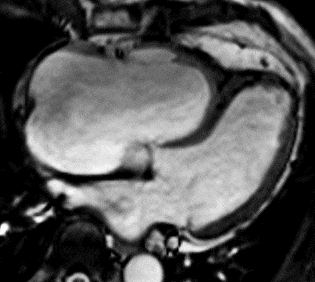
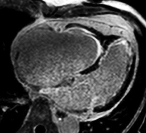
Cine and LGE 4CH: Left ventricle with particular impaired longitudinal function. Volume overloaded right ventricle with impaired systolic function and diastolic septal shift to the left due to free tricuspid regurgitation. Severely dilated atria. LGE imaging demonstrated endomyocardial pattern of fibrosis in both ventricles at the apex („V sign“). Combination of restrictive cardiomyopathy due to late stage endomyocardial fibrosis and volume overloaded right ventricle due to free tricuspid regurgitation.
|