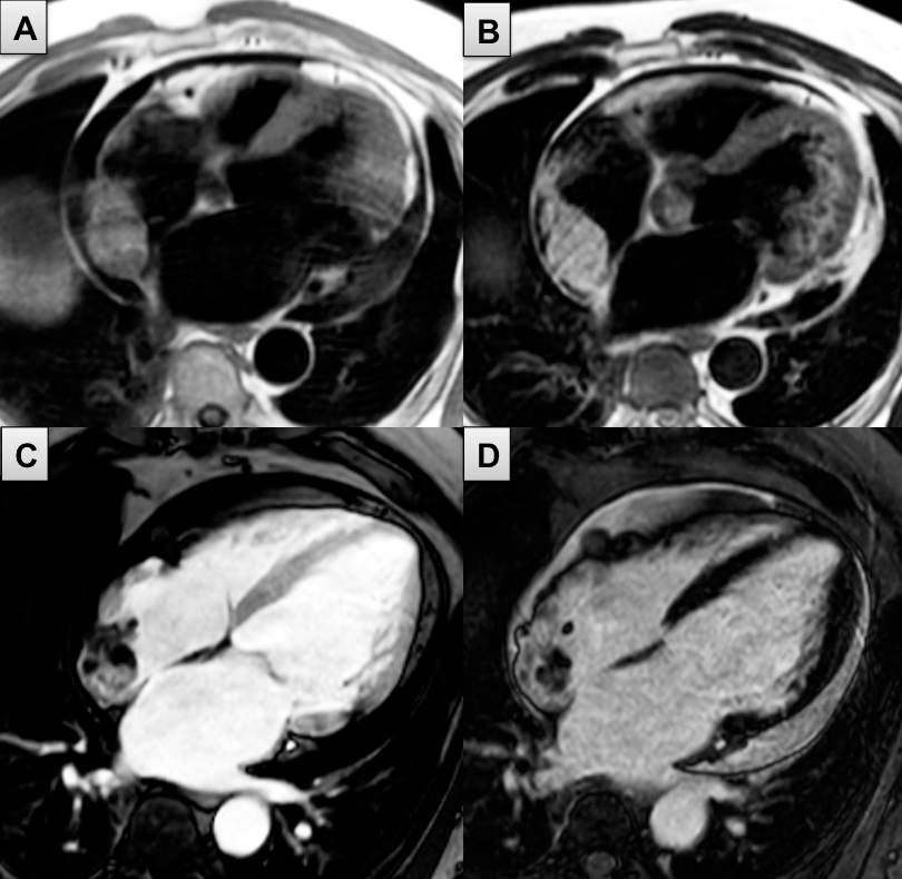
Tumour characterisation: (A) T1w 4CH: low signal intensity compared to the myocardium; (B) T2w 4CH: high signal intensity; (C) EGE 4CH: heterogeneous; (D) LGE 4CH: heterogeneous.
 
Tumour with an irregular border at the posterior wall of the RA. Global pericardial effusion not compromising RA/RV yet.

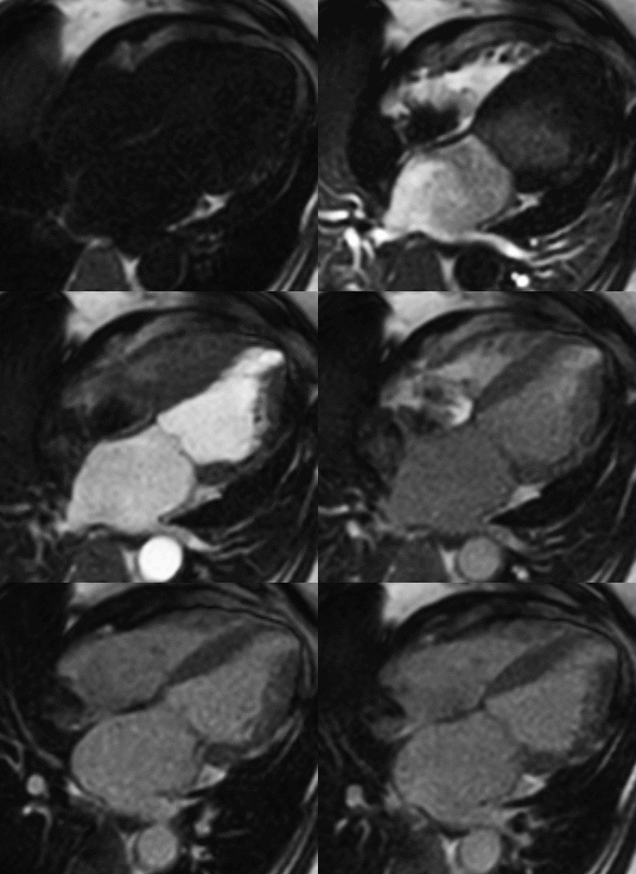
First-pass perfusion: heterogeneous nature of flow of contrast suggesting vascularity within the tumour.
|


