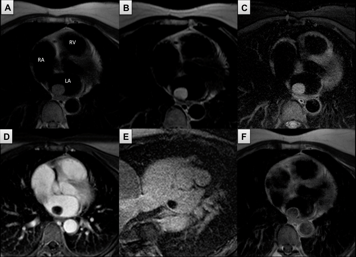
Tumour characterisation: (A) T1w SA: isointense compared to the myocardium; (B) T2w SA w/o fat suppression: high signal intensity; (C) T2w SA with fat suppression: high signal intensity; (D) EGE SA: dark; (E) LGE SA: dark with some heterogeneous area; (F) T1w post contrast: heterogeneous.
2CH cine: Left atrial round mobile mass with a stalk to the atrial wall.
3) First-pass perfusion: Minimal uptake by the LA mass.
|