Protocol
  1. High resolution anatomy module (A)
  2. Cine imaging (B) in all standard and targeted planes
  3. Consider Tagging module (J)
  4. In 2 optimized orthogonal planes:
    • T1w black blood images with and without fat suppression
    • T1w black blood images pre and post contrast
    • T2w
    • Perfusion module (D)
    • EGE and LGE module (E)
    * optional.
    + The second bolus of gadolinium-based contrast material is given with the first-pass perfusion sequence, if performed, or alternatively as a top-up dose prior to EGE imaging.
    See Reference (8).
Tissue characteristics


Modified from reference (8). * T1w and T2w imaging signal is given relative to myocardium; † best seen on early gadolinium enhancement imaging (no uptake) 2 minutes after contrast (Figure 1); ‡ similar to surrounding fat signal and characterized by marked suppression with fat-saturation pre-pulse. § the exception is metastatic melanoma which has a high T1w and a low T2w signal.
Report
  1. Location and 3 dimensional size
  2. Relation to peri-/ myocardium, valves and chamber
  3. Signal intensity on T1, T1 fat sat, T2 and STIR images
    • Homogenous or heterogeneous
    • Hyper-/ iso- / hypointense to myocardium or chest wall
  4. Margins: smooth, irregular, infiltrating, pediculated
  5. Specify motion with myocardium / pericardium
  6. Presence and location of LGE
  7. Presence of effusion (pericardial or pleural)
Key Points
  1. Cardiac metastatic lesions are up to 1000 times more common than primary tumours
  2. Common sources of metastic lesions
    • Melanoma, thyroid cancer, breast cancer, renal carcinoma, soft tissue carcinoma, lung cancer, esophageal cancer, hepatocellular carcinoma
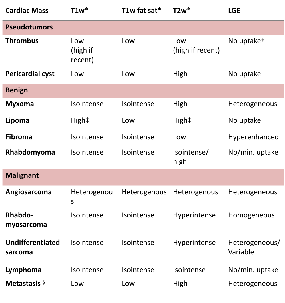
  3. Common benign primary tumours (70%)
    • Myxoma, lipoma, fibroelastoma, fibroma, rhabdomyoma, hemangioma
  4. Common malignant primary tumours (30%)
    • Angiosarcoma, rhabdomyosarcoma, mesothelioma, fibrosarcoma, lymphoma
  5. Typical tumour locations*:
    • Any chamber: Lipoma (intramural / intracavitary ), Hemangioma (intracavitary), Rhabdomyosarcoma (intramural, metastases).
    • Ventricles: Fibroma (intramural), Rhybdomyoma (intramural).
    • Valvular: Fibroelastoma, Vegetations.
    • Left Atrium: Thrombus, Myxoma, Fibrosarcoma, Osteosarcoma, Leiomyosarcoma (posterior wall), undiffertiated sarcoma.
    • Right Atrium: Angiosarcoma, Lymphoma.
    • Pericardium: Pericardial cyst, metastases.
  6. Consider pseudo tumours:
    • Normal heart structures, thrombus, cyst or vegetation

Tips and Tricks
  1. Very small and highly mobile masses (e.g. vegetation, fibroelastoma) might be missed with CMR
  2. CMR allows tissue characterisation, but cannot provide histopathologic information.