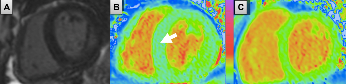
Case 1: Anderson Fabry Disease
A) LGE SA: late enhancement basal inferolateral and inferior. B) Native T1 map SA of AFD patient: low septal T1 (780 ms) appearing more blue (white arrow). C) Native T1 map SA of a healthy volunteer: normal septal T1 (968 ms) appearing more green.