Protocol
  1. Anatomy module (A)
  2. LV function (RV function) module (B/C)
  3. Edema module (G)
  4. EGE/LGE module (E)
  5. T1 Mapping module (K, if available)
Report
  1. Dimensions, mass (corrected for BSA) and function
    • LV: EDV, ESV , SV, EF, longitudinal function, mass
    • RV: EDV, ESV, SV, EF, longitudinal function
    • Regional wall motion abnormalities
    • Thickness of interatrial septum
  2. Valve regurgitation
  3. LGE pattern
  4. Pericardial / pleural effusion
Key Points
  1. Restrictive LV pattern (non-dilated ventricles, preserved LV function, restrictive filling pattern, enlarged LA / RA) and global LV hypertrophy
  2. LGE
    • Consider amyloidosis if myocardial nulling difficult to achieve on LGE images despite good technique
    • Predominantly global sub-endocardial distribution
    • Other patterns such as patchy subendocardial or transmural enhancement can also occur
    • On Look-Locker images the blood pool and myocardium cross zero (appear black) at similar timepoints
    • Cardiac involvement without any LGE is rare
  3. Atrial septum hypertrophy of >6mm (20% of cases)
  4. Pericardial and pleural effusion are common
Tips and Tricks
  1. Cross check skeletal muscle signal intensity while choosing optimal inversion time
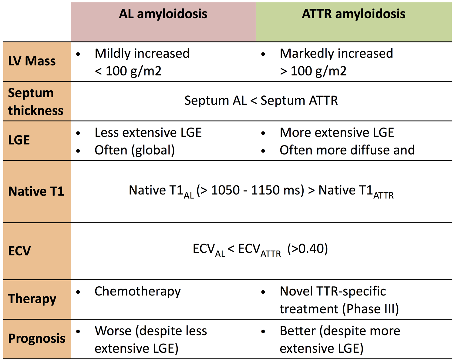
  2. For differential diagnosis AL amyloidosis vs. ATTR amyloidosis see table below


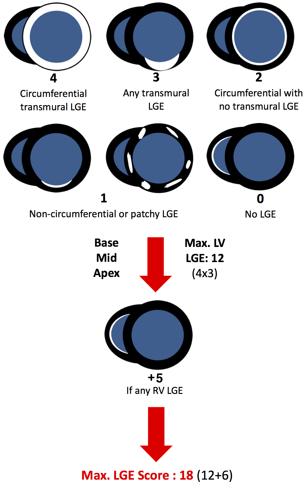
QALE score for DD AL and ATTR


References
  1. ESC Guidelines for the diagnosis and treatment of acute and chronic heart failure 2012. ESC Clinical Practice Guidelines 2012
  2. Dungu JN, Valencia O, Pinney JH, et al. CMR-based differentiation of AL and ATTR cardiac amyloidosis. JACC Cardiovasc Imaging. 2014 Feb;7(2):133-42.